

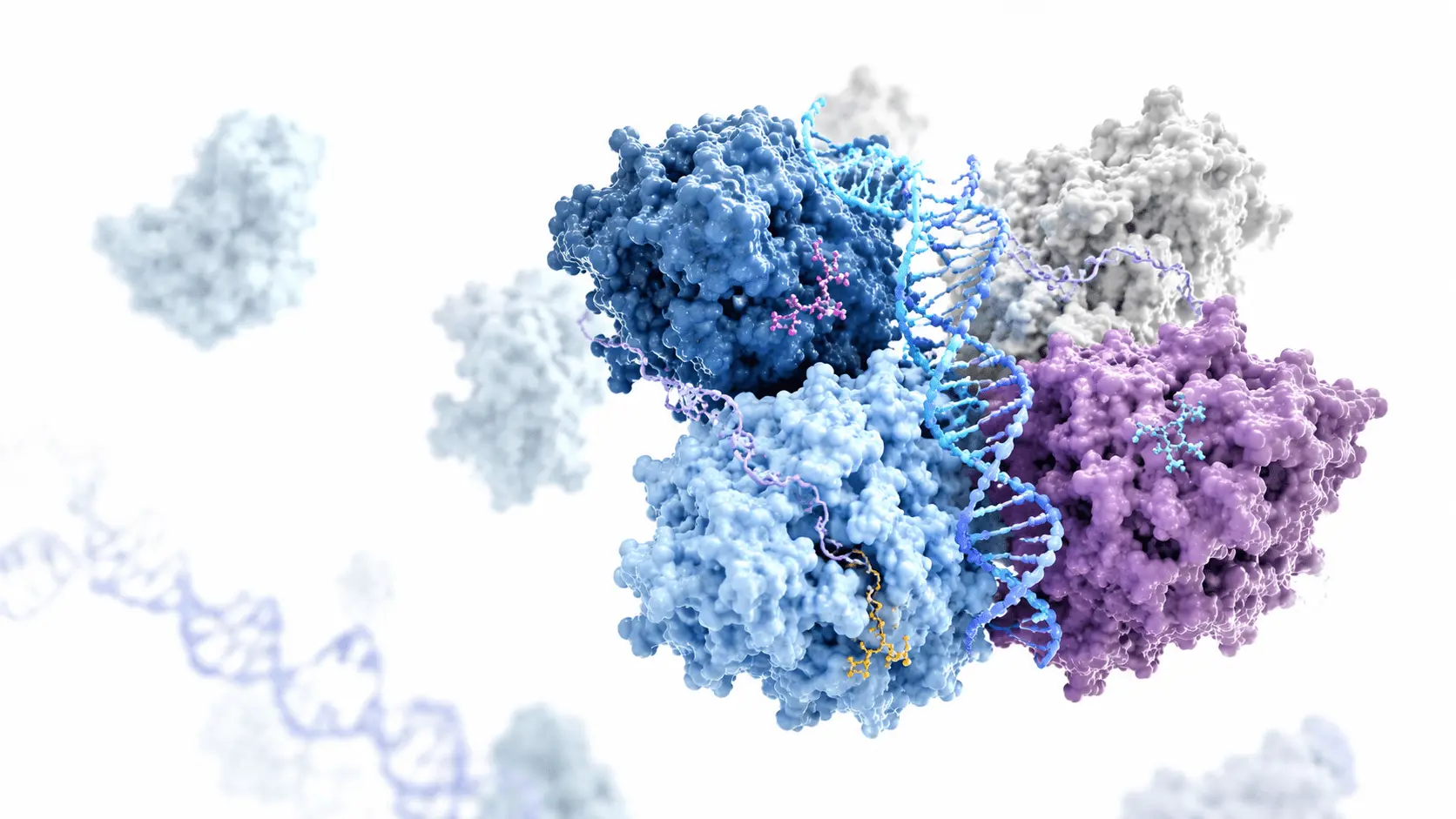
Proteins are the workers of the cell, undertaking countless tasks from enzymes to antibodies, from transporters to signaling molecules. However, fully understanding a protein's function is not possible by knowing only its amino acid sequence. Function is acquired through three-dimensional folding, and the disruption of this folding underlies many diseases, from Alzheimer's to cancer. Structural biology maps this three-dimensional world, investigating how proteins work, how they interact, and how they can be controlled.
For bioinformaticians, this field offers the opportunity to transform massive datasets into meaningful models. For chemists, visualizing molecular-level interactions serves as a guide in the design of new compounds. In this article, we will address the fundamental concepts of structural biology, the hierarchy of protein structure, and the advanced analytical techniques used today. Our goal is to present both the experimental and computational aspects of this discipline within a complementary framework.
The history of determining protein structure began in the 1950s with the solving of the crystal structure of myoglobin. Since then, experimental methods have advanced, and computational predictions have matured. Today, algorithms exist that can predict a protein's structure within seconds. However, the reliability of these predictions remains limited unless validated by experimental data. Throughout this article, we will see how these two approaches intertwine and why both are critical.
The Language of Folding: Primary, Secondary, Tertiary, and Quaternary Structure
Protein structure is examined at four distinct levels of organization. The primary structure is the linear sequence of amino acids, determined by the genetic code. This sequence forms the basis for the protein's final folding. The secondary structure consists of regular patterns such as alpha helices and beta sheets that form in local regions. These patterns are stabilized by hydrogen bonds between backbone atoms and contribute to the protein's overall shape.
Tertiary structure refers to the three-dimensional folding of an entire polypeptide chain. At this level, hydrophobic interactions, ionic bonds, and disulfide bridges between side chains play a decisive role. Quaternary structure describes the complexes formed by the assembly of multiple subunits. For example, hemoglobin consists of four subunits, and this assembly enhances its oxygen-carrying capacity.
Protein folding is a thermodynamic equilibrium process. Chaperones intervene in the cell to ensure correct folding, and misfolding triggers cellular stress responses.
Understanding these four levels is essential for comprehending a protein's function. For instance, an enzyme's active site is a result of its tertiary structure and requires a specific geometry for substrate binding. Mutations can disrupt this structure, leading to loss of function. Therefore, structural biology is also a critical tool for predicting the phenotypic effects of genetic variants.
Experimental Techniques: Crystallography, NMR, and Cryo-EM
There are three main experimental methods for determining protein structure: X-ray crystallography, nuclear magnetic resonance (NMR) spectroscopy, and cryo-electron microscopy (cryo-EM). Crystallography requires the protein to be crystallized, and an electron density map is derived from the diffraction pattern of X-rays. This method provides high resolution, but the crystallization step can be challenging and may remove the protein from its natural environment.
NMR spectroscopy examines proteins in solution and provides dynamic information. This allows transitions between different conformations of the protein to be observed. However, NMR is limited for large proteins due to signal overlap. Cryo-EM, on the other hand, preserves proteins in their native state by freezing them and enables high-resolution imaging of large complexes. In recent years, the resolution revolution in cryo-EM has expanded the boundaries of structural biology.
Cryo-EM allows protein complexes to be studied in their native states. This technique has revolutionized the study of difficult-to-crystallize samples, such as membrane proteins.
Each method has its advantages and limitations. Bioinformaticians process this experimental data to build atomic models and prepare them for molecular dynamics simulations. Chemists use these models to identify ligand-binding sites and develop targeted strategies in drug design.
Computational Approaches: Homology Modeling and Molecular Dynamics
Experimental methods are not applicable to every protein. This is where computational approaches come into play. Homology modeling predicts the structure of a protein with a similar sequence based on a known structure. This method relies on the structural similarity of evolutionarily conserved regions and yields quick results. However, if sequence similarity is low, the reliability of the prediction decreases.
Molecular dynamics (MD) simulations reveal the dynamic behavior of proteins by modeling their atomic-level movements over time. These simulations are used to study protein folding, ligand binding, and conformational changes. When combined with experimental data, MD becomes a powerful tool for understanding the mechanical basis of protein function.
- Homology modeling is widely used in genome projects to predict the function of newly sequenced proteins.
- Molecular dynamics is critical for calculating the binding affinity of drug candidates and optimizing protein stability.
- Deep learning-based approaches have revolutionized structure prediction; however, experimental validation remains the gold standard.
These computational tools are part of the daily workflow for bioinformaticians. Chemists, meanwhile, combine data from these simulations with calculations of chemical reactivity and binding energy to obtain more realistic models.
Why Structural Biology Matters: Diseases and Drug Development
Knowledge of protein structure plays a central role in elucidating the molecular mechanisms of diseases and developing new therapeutic strategies. For example, sickle cell anemia results from a single amino acid mutation in the beta chain of hemoglobin. This mutation alters the protein's folding, leading to fibril formation and distorting the shape of red blood cells. Structural biology clarifies disease mechanisms by visualizing the effects of such mutations.
In drug development, the three-dimensional structure of a target protein serves as a map for designing drug molecules. Inhibitors that bind to an enzyme's active site are optimized using structural information. Furthermore, the structural basis of protein-protein interactions enables the development of new drug classes that target these interactions.
Structural biology encompasses not only proteins but also nucleic acids and lipids. The interaction networks of these molecules provide a holistic understanding of cellular processes.
For bioinformaticians, structural data is used to predict the pathogenicity of genomic variants. Chemists aim to develop more selective and less toxic compounds through structure-based drug design. This interdisciplinary collaboration is paving the way for personalized medicine.
Future Directions: Artificial Intelligence and Integrated Approaches
Structural biology is rapidly evolving with technological advancements. Artificial intelligence-based structure prediction methods have reduced the need for experimental structure determination for many proteins. However, the training data for these models relies on experimental structures, and thus the importance of experimental methods has not diminished. On the contrary, the integration of experimental and computational methods is producing more reliable results.
In the future, real-time imaging of intracellular structures and monitoring their dynamics may become possible. This will open new horizons for understanding protein functions in their natural environments. Additionally, artificial intelligence models could accelerate systems biology studies by predicting protein-protein interaction networks.
- Structural biology can reveal cellular heterogeneity through single-molecule techniques.
- Integrated methods combine the accuracy of experimental data with the predictive power of computational models.
Knowing a protein's structure is a powerful starting point for understanding its function. However, the real challenge lies in grasping the dynamic nature of this structure and its cellular context. Structural biology provides us with the tools to overcome this challenge, and each newly solved structure adds another piece to the molecular logic of life. As an interdisciplinary bridge, this field will continue to advance us in unraveling the complexity of biology.